New users enjoy 60% OFF 152,8,717 stock photos onlineNormal Human Heart Anatomy Anterior Anatomic view Human Heart Anatomy Left oblique View Human Heart Anatomy Apical 4chamber anatomy of the human heart If you click on the images, you will see them in Full Size Feel free to download and use them asA heart attack occurs when the flow of blood to the heart muscle suddenly becomes blocked If blood flow is not restored, heart muscle begins to die Learn more about causes, risk factors, screening and prevention, signs and symptoms, diagnoses, and treatments for heart attack, and how to participate in clinical trials
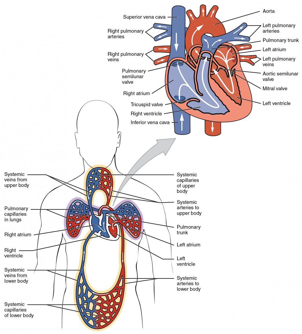
Heart Anatomy Anatomy And Physiology
Full body heart diagram
Full body heart diagram-The heart is a large muscular organ with the very important job of circulating blood through the blood vessels to the body Located in the center of the chest, the heart is the hardest working muscle in the human body — always working, even while we are sleeping The heart and blood vessels together make up the body's cardiovascular systemThe heart is a muscular organ about the size of a fist, located just behind and slightly left of the breastbone The heart pumps blood through the network of arteries and veins called the


How To Draw The Internal Structure Of The Heart With Pictures
Review how the atrioventricular and semilunar valves open and close in a full cardiac cycle in this interactive tutorial The Location, Size, and Shape of the Heart Review the location of the heart as well as it's size and shape in this interactive tutorial and test your knowledge in the quiz 3D Oblique view of the human heart;Heart Diagram Diagram of a heart Human Heart Human Heart Anatomy The human heart consists of the following parts aorta, left atrium, right atrium, left ventricle, right ventricle, veins, arteries and others Heart diagram with labels Human Body Anatomy Diagrams Primary menu Skip to primary content
The state diagram contains regional information from seven positions in the heart and global information represented by the mean time intervals The outermost circular segment in the state diagram corresponds to the anteroseptal wall, followed by the anterior, anterolateral, inferolateral, inferior, inferoseptum (LV) and medial wall (RV)Heart Diagram Body Diagram Human Body Anatomy Human Anatomy And Physiology Nursing Goals College Nursing Cardiac Anatomy Cardiac Cycle Heart Anatomy The heart pumps around 57 litres of blood in a day throughout the body The heart is situated at the centre of the chest and points slightly towards the left On average, the heart beats about 100,000 times a day, ie, around 3 billion beats in a lifetime The average male heart weighs around 280 to 340 grams (10 to 12 ounces)
The heart weighs between 7 and 15 ounces (0 to 425 grams) and is a little larger than the size of your fist By the end of a long life, a person's heart may have beat (expanded and contracted) more than 35 billion times In fact, each day, the average heart beats 100,000 times, pumping about 2,000 gallons (7,571 liters) of bloodIntroduction to Anatomy of the Heart This course is designed to give you a comprehensive introduction to the anatomy of the heart It is an interactive, lecture based course covering the underlying concepts and principles related to human gross anatomy of the heartHeart diagram The myocardium and coronary vessels;


Human Heart Diagram Labeled Science Trends
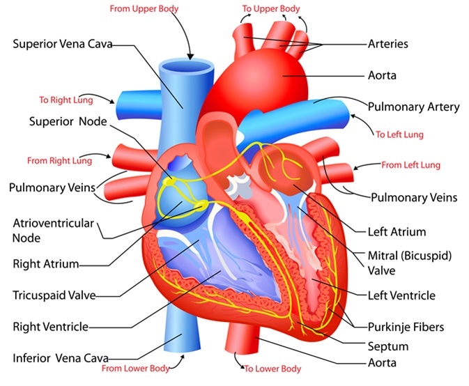


Structure And Function Of The Heart
Heart STUDY Learn Flashcards Write Spell Test PLAY Match Gravity Created by armstrong08 Terms in this set (16) Left ventricle pumps blood through the aorta to the rest of the body Right Atrium Receives deoxygenated blood from the body Right Ventricle Pumps blood to the lungs AortaHeart pumps pure blood to different parts of the body and then takes the deoxygenated blood from all the parts to the lungs for oxygenation Normally in a minute the heart beats 72 times and pumps around 1,500 to 2,000 gallons of blood per day Let's check out heart diagram which can help you to understand functioning of the heart in a better Label the heart In this interactive, you can label parts of the human heart Drag and drop the text labels onto the boxes next to the diagram Selecting or hovering over a box will highlight each area in the diagram Aorta The main artery


Heart Detail Picture Image On Medicinenet Com


Heart Cross Section Labeling Diagram Quizlet
The wall of two ventricles are strong and sturdy when compared to atria Before we start, we shall recall the basic proportions of heart and its chambers The right Auricle is larger than left The walls of Left ventricle are thicker than the walls of Right ventricleWe shall try to incorporate the above details in the diagram for maximum accuracy Quiz Identify Various Parts Of Human Heart The human heart is a vital organ for every human The more healthy your heart is, the longer the chances you have of surviving, so you better take care of it Take the following quiz to know how much you know about your heart Upgrade and get a lot more done!Hello EveryoneHuman Heart Diagram How to Draw Human Heart Diagram Step By Step Class 10 Biology NCERTHuman Heart Diagram, How to Draw Human Hear
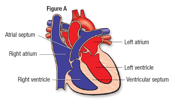


How The Healthy Heart Works American Heart Association
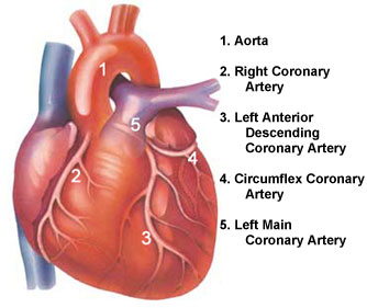


How The Heart Works Froedtert The Medical College Of Wisconsin
So what you're looking at is one of the most amazing organs in your body this is the human heart and it's shown with all the vessels on it and you can see the vessels coming into it and out of it but the heart at its core is a pump and this pump is is why we call it the hardest working organ in our body because it starts pumping blood from the point where you're a little fetus maybe aboutDownload 8,914 Diagram Heart Stock Illustrations, Vectors & Clipart for FREE or amazingly low rates!The heart is made of three layers of tissue Endocardium, the thin inner lining of the heart chambers that also forms the surface of the valves Myocardium, the thick middle layer of muscle that allows your heart chambers to contract and relax to pump blood to your body Pericardium, the sac that surrounds your heart Made of thin layers of tissue, it holds the heart in place and
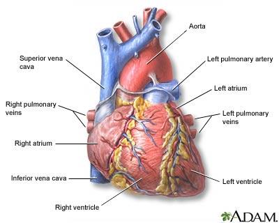


Heart Front View Medlineplus Medical Encyclopedia Image


Heart How Your Heart Pumps Blood Around Your Body Mydr Com Au
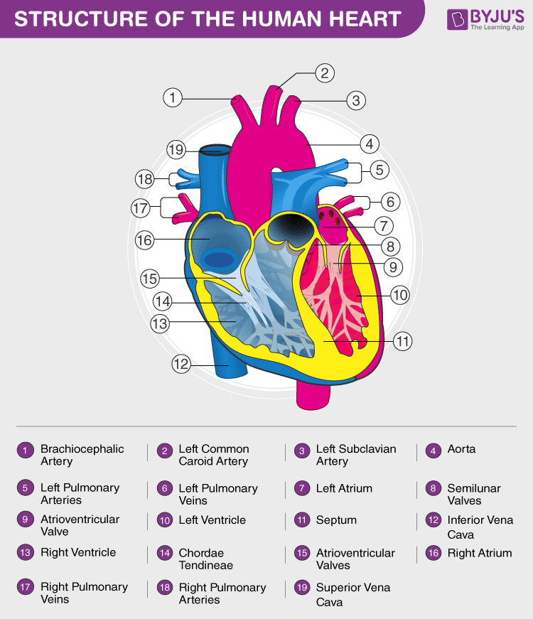
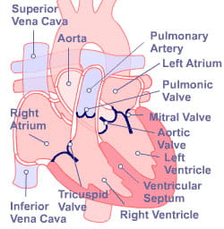
A Labeled Diagram of the Human Heart You Really Need to See The heart, one of the most significant organs in the human body, is nothing but a muscular pump which pumps blood throughout the body The human heart and its functions are truly fascinating The heart, though small in size, performs highly significant functions that sustains human lifeExplore the diagram of heart along with the structural details only at byju'sDiagram of the human heart, created by Wapcaplet in Sodipodi Cropped by Yaddah to remove white space (this cropping is not the same as Wapcaplet's original crop) English Diagram of the human heart 1 Superior vena cava 2 4 Mitral valve 5 Aortic valve 6 Left ventricle 7 Right ventricle 8 Left atrium 9 Right atrium 10 Aorta


Anatomy Of The Heart Human Heart Anatomy Heart Anatomy Gross Anatomy


Anatomy Of A Human Heart
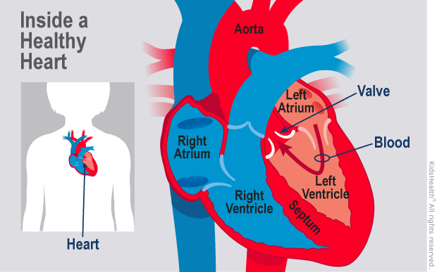
As shown in the human heart diagram above, the heart has several cavities (ventricles and atrium) and valves (pulmonary, aortic, mitral, tricuspid) which move blood inandout of the heart It's pretty amazing to watch blood flow through the heart To see how blood flows through the heart using an animation, please click hereThe heart is a muscular organ in most animals, which pumps blood through the blood vessels of the circulatory system The pumped blood carries oxygen and nutrients to the body, while carrying metabolic waste such as carbon dioxide to the lungs In humans, the heart is approximately the size of a closed fist and is located between the lungs, in the middle compartment of the chest The heart is a mostly hollow, muscular organ composed of cardiac muscles and connective tissue that acts as a pump to distribute blood throughout the body's tissues


1
(230).jpg)


Quiz Identify Various Parts Of Human Heart Proprofs Quiz
Female anatomy full body 3d rendered illustration of the female anatomy diagram of body organs female pics stock pictures, royaltyfree photos & images Human internal organs vector Vector isolated illustration of human internal organs in female body Stomach, liver, intestine, bladder, lung, testicle, uterus, spine, pancreas, kidney, heartStudents will conduct a series of investigations that model the functions and parts of the heart Students will be able to correctly annotate a diagram of the circulatory system and will identify, label and correctly place veins, arteries, capillaries and a heart diagram Vertices 971k More model information Anatomical cross section of the human heart showing the internal working and complexity of one of the hardest working organs in the body Published 3 years ago Mar 19th Art & abstract 3D Models


How Long Does It Take For Full Heart Attack Recovery
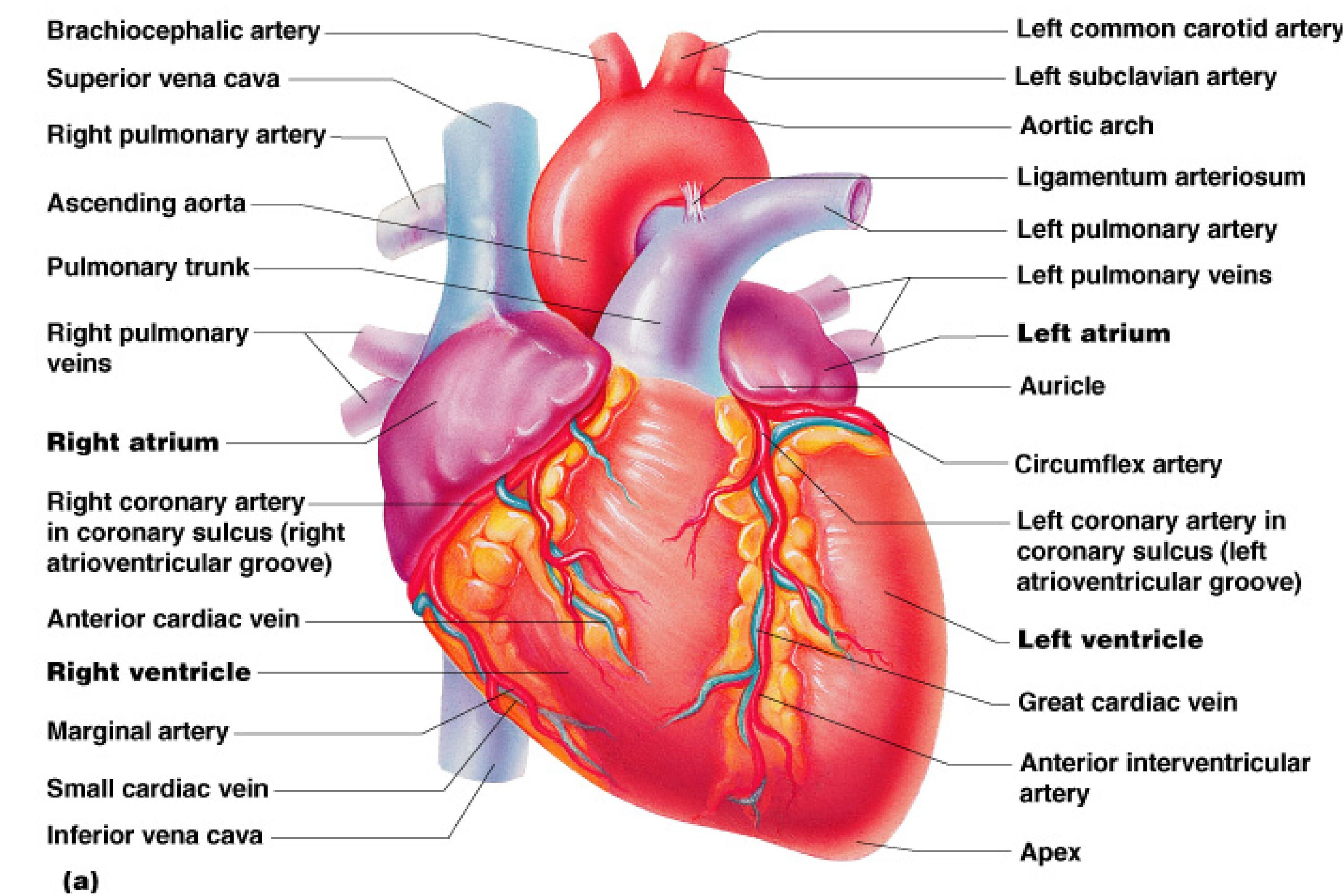


Real Heart Diagram Poster Etsy
Its pumping power also pushes blood through organs like the lungs to remove waste products like CO2;An echocardiogram is a test that uses ultrasound to show how your heart muscle and valves are working The sound waves make moving pictures of your heart so your doctor can get a Posted on by Admin 13 Full Heart Diagram Drawing human heart's diagram is very easy 600 x 600 photo description Tachycardia Causes, Symptoms and Treatments – Medical from wwwmedicalnewstodaycom How to draw human heart step by step ?


Full Heart Diagram Pt 3 Diagram Quizlet


Diagram Google Images Heart Diagram Full Version Hd Quality Heart Diagram Outletdiagram Electricdriveitalia It
Best Free png HD http //www anatomybox com/wp heart diagram human heart full diagram png images background, PNG png file easily with one click Free HD PNG images, png design and transparent background with high quality This file is all about PNG and it includes http //www anatomybox com/wp heart diagram human heart full diagram tale whichDiagram of the blood flow path in the heart Now that blood is freshly oxygenated from its trip to the lungs, it returns to the left atrium of the heart and passively flows through the bicuspid (or A heart diagram labeled will provide plenty of information about the structure of your heart, including the wall of your heart The wall of the heart has three different layers, such as the Myocardium, the Epicardium, and the Endocardium Here's more about these three layers
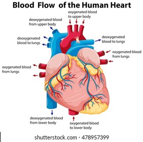


Diagram Of The Human Heart Images Stock Photos Vectors Shutterstock
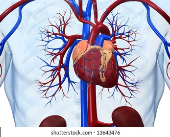


Human Heart Diagram High Res Stock Images Shutterstock
The heart is an amazing organ It starts beating about 22 days after conception and continuously pumps oxygenated red blood cells and nutrientrich blood and other compounds like platelets throughout your body to sustain the life of your organs; The cardiac cycle is the sequence of events that occurs when the heart beats As the heart beats, it circulates blood through pulmonary and systemic circuits of the body There are two phases of the cardiac cycle The diastole phase and the systole phase In the diastole phase, heart ventricles relax and the heart fills with blood Heart anatomy The heart has five surfaces base (posterior), diaphragmatic (inferior), sternocostal (anterior), and left and right pulmonary surfaces It also has several margins right, left, superior, and inferior The right margin is the small section of the right atrium that extends between the superior and inferior vena cava
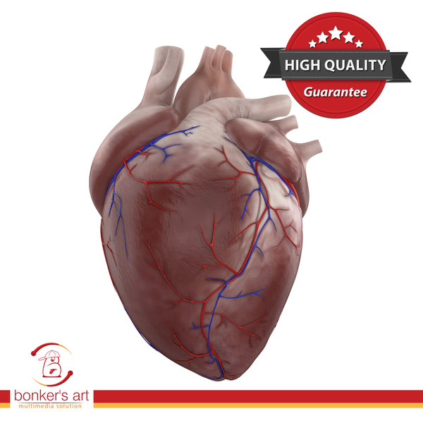


3d Human Heart Turbosquid


How To Draw The Internal Structure Of The Heart With Pictures
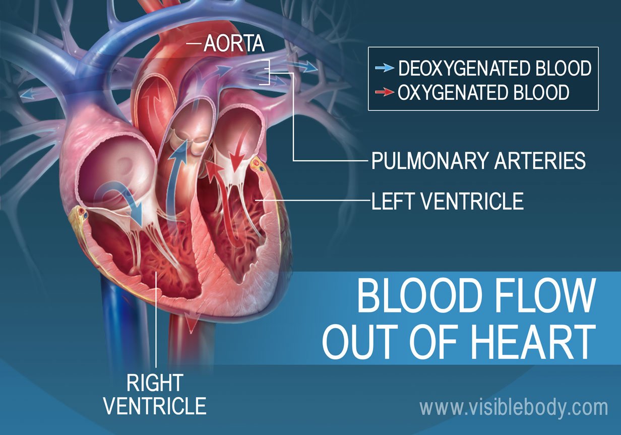
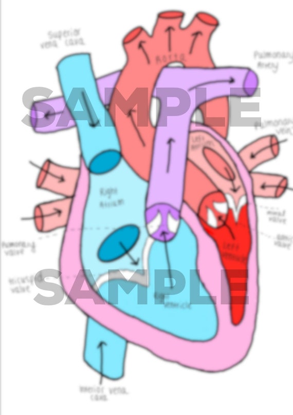
Blood enters the heart through two large veins – the posterior (inferior) and the anterior (superior) vena cava – carrying deoxygenated blood from the body into the right atrium Blood flows from the right atrium into the right ventricle through the tricuspid valve When the ventricle is full, the tricuspid valve shuts to prevent blood Identify Various Parts Of A Human Heart Trivia Quiz The heart is the most important organ in the body It is in charge of keeping the processes within the body moving by facilitating the transfer of blood throughout the body The quiz below is to test out interesting facts you may know about the heartHCL Learning DigiSchool presents you animated study material on structure of the Human Heart It describes the location, structure and function of the human
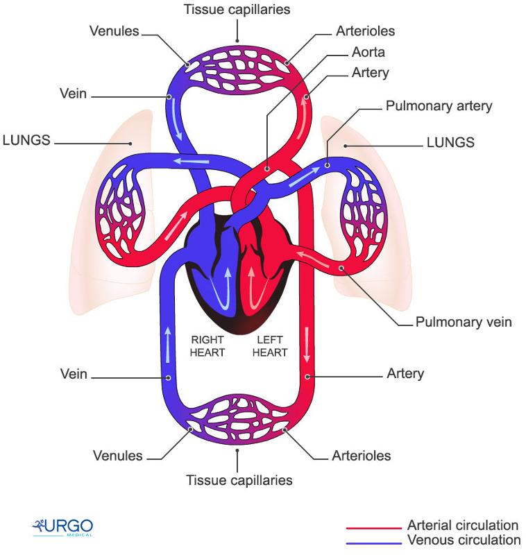


Cardiovascular System


Internal Heart Diagram Diagram Quizlet
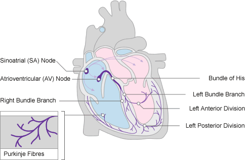
Circulatory system diagrams are visual representations of the circulatory system, also referred to as the cardiovascular system It is comprised of three parts the pulmonary circulation, coronary circulation, and systemic circulation The main function of the circulatory system is to circulate blood, which carries oxygen and nutrients The heart's electrical signal is produced by a tiny structure known as the sinus node, which is located in the upper portion of the right atrium (The anatomy of the heart's chambers and valves includes two atria at the top of the heart with two ventricles at the bottom)Shape and Size of the Heart The shape of the heart is similar to a pinecone, rather broad at the superior surface and tapering to the apex A typical heart is approximately the size of your fist 12 cm (5 in) in length, 8 cm (35 in) wide, and 6 cm (25 in) in thickness


Heart Diagram Right Left Atria Right Left Ventricles Pulmonary Trunk Aorta Superior Inferio Human Heart Anatomy Heart Anatomy Human Anatomy And Physiology


Heart Anatomy Anatomy And Physiology
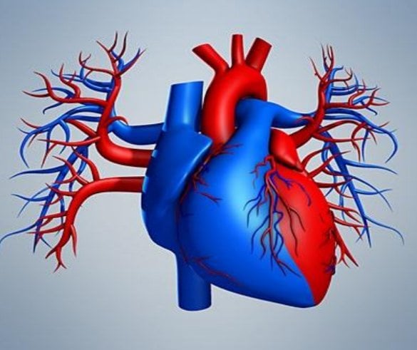
The heart is a muscular organ about the size of a closed fist that functions as the body's circulatory pump It takes in deoxygenated blood through the veins and delivers it to the lungs for oxygenation before pumping it into the various arteries (which provide oxygen and nutrients to body tissues by transporting the blood throughout the body)
:max_bytes(150000):strip_icc()/GettyImages-626974567-defc9220866c4e679c8d244dfbb997bb.jpg)


How Blood Flows Through The Heart And Lungs


Heart Structure Function Diagram Anatomy Facts Britannica


Human Heart Labeled Diagram The Human Heart Diagram Labeled Human Anatomy Photo Human Heart Labeled Diagram Th Heart Diagram Human Heart Diagram Human Heart


Know Your Heart Oxygenated And Deoxygenated Blood Heart Diagram Full Size Png Download Seekpng


Heart Anatomy Labeled Diagram Structures Blood Flow Function Of Cardiac System Ezmed


Heart Wikipedia


The Living Heart Project A Robust And Integrative Simulator For Human Heart Function Sciencedirect


Diagram Human Heart Diagram Unlabeled Full Version Hd Quality Diagram Unlabeled Hpvdiagrams Albergotrepozzi It


Human Heart Diagram Royalty Free Cliparts Vectors And Stock Illustration Image


File Heart Diagram En Svg Wikipedia
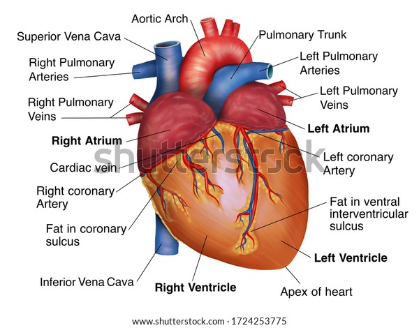


Heart Anatomy Full Part Study Education Stock Illustration


Male Full Body Circulatory System Highlights Heart Stock Photo Download Image Now Istock
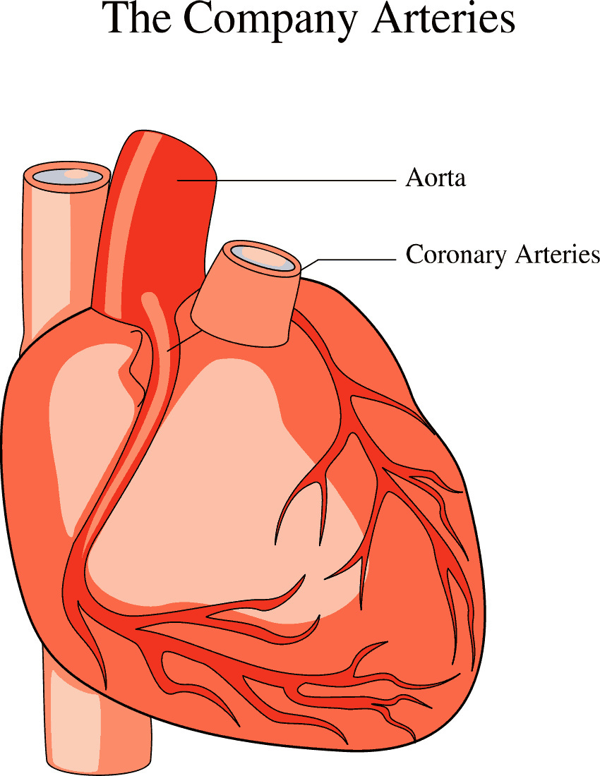


Heart Diagram Full Page
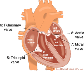


How Artificial Hearts Work Howstuffworks


How The Heart Works Froedtert The Medical College Of Wisconsin


Diagram Of The Micro Spherical Heart Pump Shown In A A Full View Download Scientific Diagram
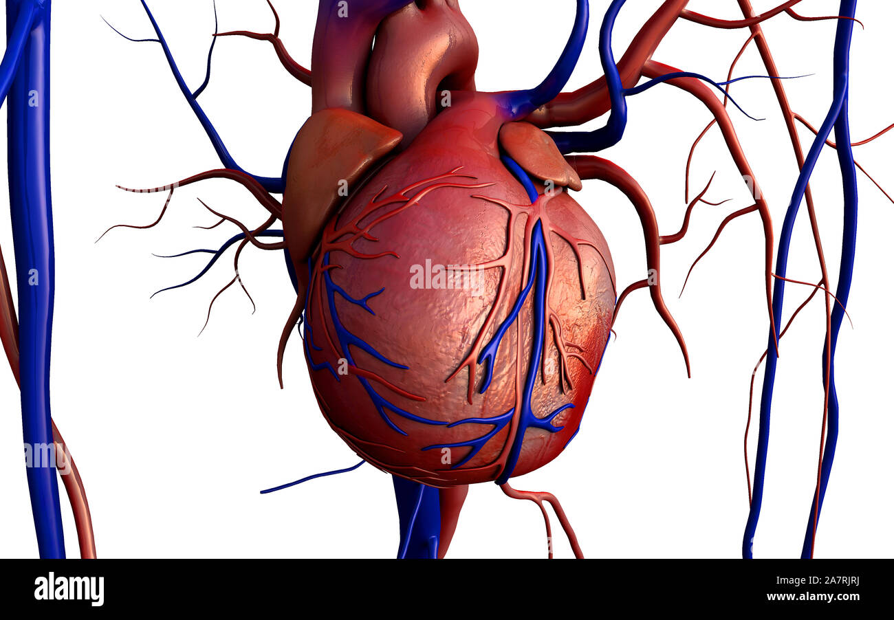


Heart Model Heart Full Clipping Path Included Human Heart For Medical Study Human Heart Anatomy Stock Photo Alamy
:background_color(FFFFFF):format(jpeg)/images/library/11110/Heart_Thumbnail.png)


Heart Anatomy Structure Valves Coronary Vessels Kenhub


Sponsored Blog S Heart Diagram A Level


Left Side Of Heart Diagram Of A Cow Heart Clipart Full Size Clipart Pinclipart


Show Me A Diagram Of The Human Heart Here Are A Bunch


Heart Model Heart Full Clipping Path Included Human Heart For Medical Study Human Heart Anatomy Stock Photo Alamy


Transverse Section Of Human Heart 5 Download Scientific Diagram
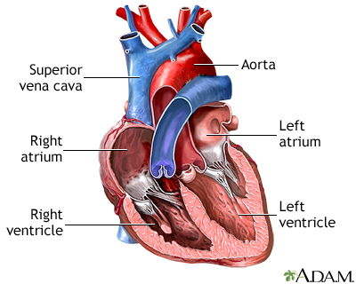


Heart Chambers Medlineplus Medical Encyclopedia Image


The Living Heart Project A Robust And Integrative Simulator For Human Heart Function Sciencedirect
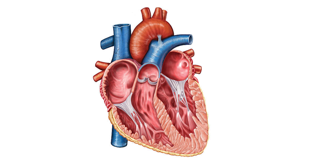


Heart Anatomy Labeled Diagram Structures Blood Flow Function Of Cardiac System Ezmed


Heart Anatomy Anatomy And Physiology


1
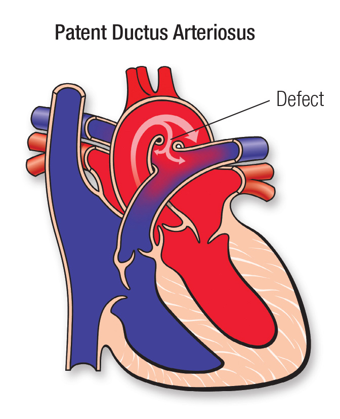


Patent Ductus Arteriosus Pda American Heart Association
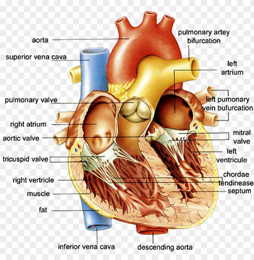


Http Www Anatomybox Com Wp Heart Diagram Human Heart Full Diagram Png Image With Transparent Background Toppng
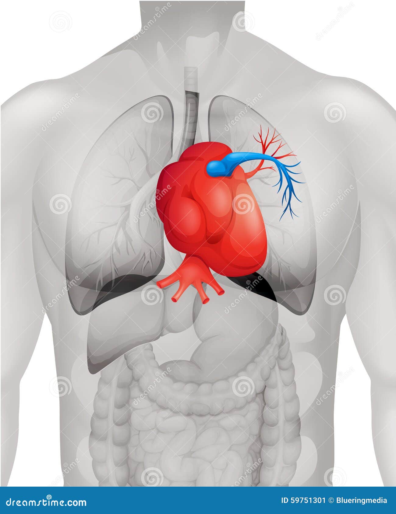


Human Heart Diagram In Detail Stock Vector Illustration Of Health White


Heart Anatomy Yourheartvalve


How The Heart Works Diagram Anatomy Blood Flow


Diagram Of Heart How To Draw Human Heart Easily Human Heart Human Heart Diagram Labelled Human Heart Youtube
/human-heart-circulatory-system-598167278-5c48d4d2c9e77c0001a577d4.jpg)


Av And Semilunar Heart Valves


File Diagram Of The Human Heart Cropped Svg Wikipedia


Physiology Tutorial The Human Heart


Seer Training Structure Of The Heart


Heart Diagram With Labels And Detailed Explanation


Heart Diagram Currituck County


Diagram Of The Human Heart Heart Of Human Full Size Png Download Seekpng


How The Heart Works Heart Foundation


The Heart Circulatory Anatomy


Heart Anatomy Anatomy And Physiology


Labelling The Heart Science Learning Hub


Cardiac Conduction System Normal Function Of The Heart Cardiology Teaching Package Practice Learning Division Of Nursing The University Of Nottingham
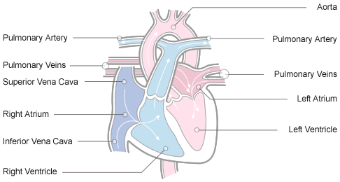


Anatomy And Physiology Of The Heart Normal Function Of The Heart Cardiology Teaching Package Practice Learning Division Of Nursing The University Of Nottingham


Diagram Label The Heart Diagram Answers Full Version Hd Quality Diagram Answers Diagramland Albergotrepozzi It
:background_color(FFFFFF):format(jpeg)/images/article/en/heart/yh35Prra0VpHLwLZLrEDA_7dpDZkx6EDgihq981V55Qg_Apex_cordis_02-2.png)


Heart Anatomy Structure Valves Coronary Vessels Kenhub
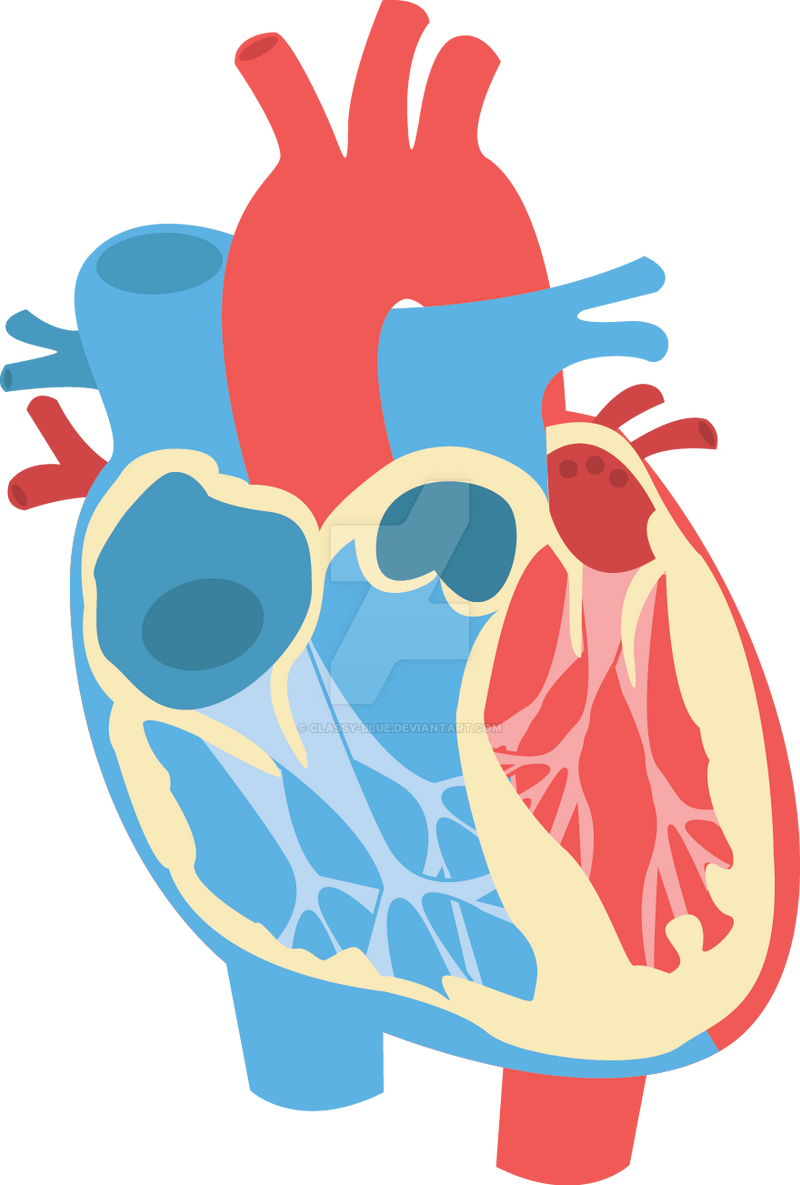


Human Heart Diagram By Classy Blue On Deviantart


Your Heart Blood Vessels
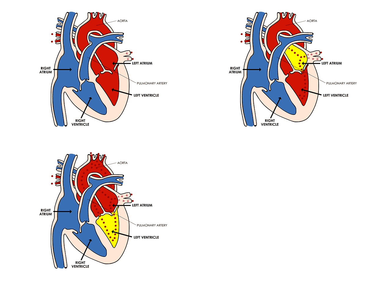


Heart And Circulatory System
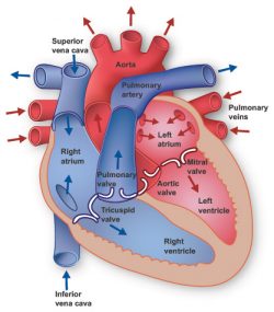


Heart Information Center Heart Anatomy Texas Heart Institute


Pin On Anatomy And Physiology


Pin On Health


Free Human Heart Sketch Diagram Download Free Human Heart Sketch Diagram Png Images Free Cliparts On Clipart Library


Anatomy Of The Heart Anatomical Chart Anatomical Chart Com Anatomy Amazon Canada


Human Heart Anatomy Functions And Facts About Heart


1


1


Heart And Circulatory System For Teens Nemours Kidshealth


Heart Structure Function Diagram Anatomy Facts Britannica


Heart Structure Function Diagram Anatomy Facts Britannica


How Your Heart Works


Anatomy Of The Human Heart Printable 6th 12th Grade Teachervision


Heart Components


File Blausen 0457 Heart Sectionalanatomy Png Wikimedia Commons


Circulation Of Heart Template Labeled Diagram Etsy


96 175 Human Heart Stock Photos Pictures Royalty Free Images Istock
/heart_inner_section-577d5c673df78cb62c939314.jpg)


Atria Of The Heart Function


Three Chambered Heart Definition Anatomy Video Lesson Transcript Study Com


13 Heart Diagram Templates Sample Example Format Download Free Premium Templates
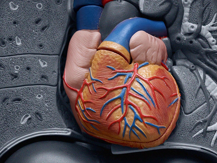


Where Is The Heart Located In The Body And How Does It Work


Video Heart And Circulatory System How They Work Mayo Clinic
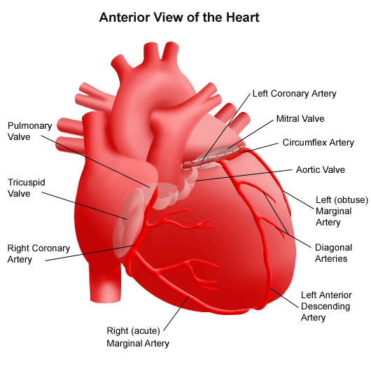


About The Heart And Blood Vessels


Baby Leino The Miracle Of Life
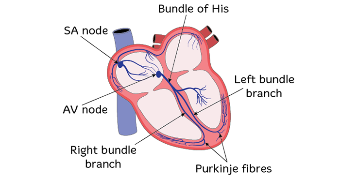


How The Heart Works Heart Foundation


Human Heart Full Explained In Hindi In Just 5 Minutes Working Or Function Youtube
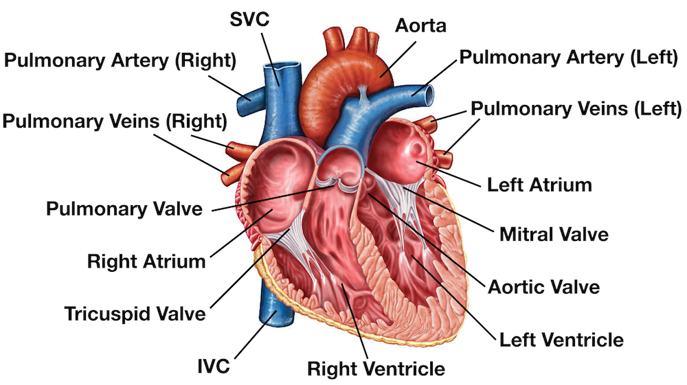


Heart Anatomy Labeled Diagram Structures Blood Flow Function Of Cardiac System Ezmed
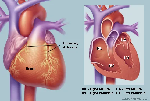


Human Heart Anatomy Diagram Function Chambers Location In Body


0 件のコメント:
コメントを投稿